"Over 40 Years In Practice;
Accredited by the American College of Radiology (ACR) Since 1998"
Porter Radiation Oncology
3663 Bee Ridge Road
Sarasota, FL 34233
Procedures / Services
Procedures / Services
IMRT
MRT involves varying (or modulating) the intensity of the radiation being used as the therapy for cancer. It is a new form of radiation therapy that uses computer-generated images to plan and then deliver more tightly focused radiation beams to cancerous tumors than is possible with conventional radiotherapy. With this capability, physicians can deliver a precise radiation dose that conforms to the shape of the tumor, while significantly reducing the amount of radiation to surrounding healthy tissues. Consequently, the technique can increase the rate of tumor control while significantly reducing side effects.
MRT uses computer-generated images to plan and then deliver tightly focused radiation beams to cancerous tumors. Physicians use it to exquisitely "paint" the tumor with a precise radiation beam that conforms as closely as possible to the shape of the tumor.
IMRT can be used to treat tumors that might have been considered untreatable in the past due to close proximity of vital organs and structures. Treating such tumors requires tremendous accuracy. In the case of head and neck tumors, IMRT allows radiation to be delivered in a way that minimizes exposure of the spinal cord, optic nerve, salivary glands, or other important structures. In the case of prostate cancer, exposure or the nearby bladder or rectum can be minimized. IMRT is being used to treat tumors in the brain, breast, head and neck, liver, lung, nasopharynx, pancreas, prostate, and uterus.
A powerful computer program optimizes a treatment plan based on a physician's dose instructions, and information about the tumor size, shape, and location in the body. A medical linear accelerator, equipped with a special device called a multileaf collimator that shapes the radiation beam, delivers the radiation in accordance with the treatment plan. The equipment can be rotated around the patient to send radiation beams from the most favorable angles for giving the tumor a high dose while preserving important healthy tissues.

External Beam Radiation Therapy
Most patients receiving treatment with radiation for cancer will be treated with external beam radiation therapy. This involves directing a beam of high energy radiation at a specific site of the body. The machine most often utilized for this treatment is a linear accelerator. This machine is capable of producing both high energy photons (X-rays) and an electron beam. The photon beam is used to treat tumors within the body, while the electron beam is used to treat more superficial areas. Some patients will receive treatment with both types of radiation.
The radiation beam is directed to a specific area using a light field, lasers, and measurements from specific points to
3D Conformal Radiation Therapy
This represents state of the art radiation therapy treatment planning. Patients treated in this manner are simulated for their treatment using a dedicated ct scanner. Data from this scan is downloaded directly into the treatment planning computer. The Radiation Oncologist will define the tumor volume and the normal structures of interest on each ct image. These images are then used to construct a three dimensional model within the computer of the relevant anatomy. The radiation treatment beams are then constructed with the aid of these images to optimally treat the cancerous area with a minimum of treatment to the surrounding tissues.

Prostate Seed Implants
Our prostate brachytherapy (seed implant) program was developed in 1990 by Dr. Galloway after he received his training in the procedure from Drs. Blasko, Grimm and Ragde at the Pacific Northwest Cancer Institute in Seattle. The program has evolved and has remained current with new developments over the years. We have always utilized the latest technology with state of the art equipment currently including Varian MMS computer planning, dedicated B & K Leopard ultrasound, and Barzell-Whitmore stabilizing stand.
The procedure is done with out-patient pre-planning of

High Dose Rate Brachytherapy
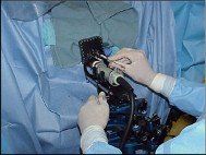
This very specialized form of radiation treatment involves the placement of a very small radioactive source of Iridium into the center of a tumor. This is often done by placing a small catheter into the area of interest using either bronchoscopy or endoscopy. Because these procedures involve certain risks, our group has collaborated with Sarasota Memorial Hospital to provide this treatment within the hospital complex.
The HDR suite is actually a small operating room and the procedure may by performed there. The placement of a catheter may be done in the endoscopy suite with

Intracavitary Low Dose Rate Brachytherapy
This form of radiation treatment is usually associated with the treatment of gynecological cancers such as cancer of the uterus or cervix. The procedure begins with the insertion of devices to hold radioactive Cesium sources within the uterus or vagina. This is generally done under general anesthesia in the operating room. After the device is properly positioned the patient is returned to a private hospital room. Later in the day radioactive Cesium sources are inserted into the delivery device. The system usually remains in place for several days, slowly delivering radiation to the interior of the tumor. In this manner a high dose of radiation is delivered to the tumor. Most patients will also receive external beam radiation as a part of their treatment.

Eye Implants for Choroidal Melanoma
The brachytherapy program for treatment of malignant melanomas involving the eye was started by Dr. Dickens in 1988 after he visited the Hahnemann University Department of Radiation Oncology and the Wills Eye Hospital in Philadelphia, Pennsylvania where this procedure underwent significant development. The procedure is done in cooperation with an experienced ophthalmologist (retinal surgeon) and a radiation physicist. The purpose of the procedure is an attempt treat the melanoma while maintaining sight in the eye and thus avoiding the necessity of surgical removal of the eye.

Therapeutic Nuclear Medicine
A wide variety of problems can be treated with the administration of radioactive isotopes either by mouth or by vein. This rapidly expanding field of medicine involves giving a radioactive substance to the patient and allowing the isotope to deliver a therapeutic dose of radiation to a target organ by concentrating the uptake of the substance within the target organ. An example would be the use of I-131 to treat an overactive thyroid gland or thyroid cancer.
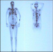
We are able to use other isotopes to treat breast or prostate cancer that has spread to bone. Exciting new developments will soon include the use of radioactive monoclonal antibodies for the treatment of certain lymphomas. These treatments are generally given in an out patient setting and take only minutes to administer. Certain precautions after treatment may be necessary with respect to contact with other people, depending upon the dose administered.

Tomotherapy
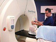
A technological breakthrough in personalized cancer treatment has "Beamed-On" at Porter Radiation Oncology, Bee Ridge Ridge, Sarasota.
The next generation radiation therapy treatment, the TOMOTHERAPY HY-ART Integrated CT-Scan/Accelerator, has arrived.
The cancer patient receiving TOMOTHERAPY begins each treatment day with a CT-Scan taken inside the TOMOTHERAPY unit. This scan shows the exact size, shape and location of the cancerous tumor and directs an x-ray beam to the precise tumor location. TOMOTHERAPY slowly "carries" the patient past thousands of small, focused radiation beams that bombard the tumor during a spiral, 360 degree pattern.
RESULTS: Doctors can safely deliver a higher radiation dose per treatment to increase curability and reduce treatment days.
Daily pre-treatment CT-Scans with instant computer adjustment of treatment beams reduces doses to surrounding normal tissue, thereby decreasing side effects and "TOMOTHERAPY is ideal for prostate cancers that typically change position daily due to changes in urine volume in the bladder," says Alan Porter, MD, Director.
The Helical (spiral) 360 degree x-ray treatment is the ultimate IMRT/IGRT system which daily adapts to tumor movement and changing shapes, TOMOTHERAPY treats tumors of the brain, lung, spinal cord, liver, pelvic area, and the head and neck. Body and brain radiosurgery (SRS, SRBT) patients are ideal candidates who may require daily treatments for only one to five days.


©2015 Dex Media. All rights reserved
Powered by Dex Media


